Shih Lab, University of Michigan,
As a part of the SURE undergraduate summer research program, I worked in the University of Michigan’s lab in biomedical manufacturing under professor Albert Shih. My research focused on making phantoms to mimic rat brain behavior during microelectrode insertion and creating skullcaps to hold probes in rat brains over long periods.
Published conference paper on custom skull caps(pdf)
Rat Brain Phantoms
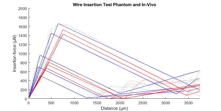
When designing microelectrodes to insert into brains for monitoring signals, you want probes that are thin and soft so the brain tissue does not identify it as foreign at attack it. However, when making a probe this pliable, it may buckle upon contact with the brain and be unable to enter. Currently the industry tests their electrodes on actual rats or agar that only simulates the brain compression behavior but my research focused on making the worlds first phantom to accurately model the insertion of a microwire into a rat brain.

My phantoms are made from PVC plastic. By changing the ratio of polymer and softener in the solution, I can adjust the hardness of each phantom. I then conducted factorial tests to change each parameter, see how each one affects various parts of the insertion plot such as slope or penetration force, and generate regression equations to predict the behavior given any quantity of each compound. Using the results from previous in-vivo tests on a rat brain, I created phantoms that closely mimicked the actual behavior and a procedure to make phantoms for other animals in the future.
3D Printed Rat Brain Skullcap
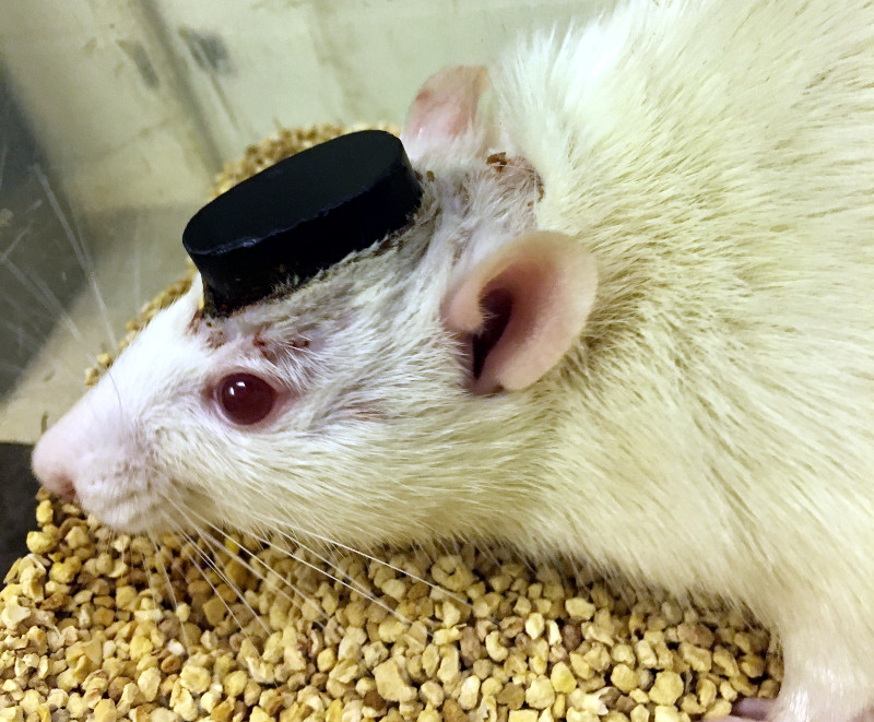
When conducting long term tests on the brain activity of rats, you need a cap that securely mounts onto the skull of the rat while holding the probes in place. Additionally, the cap helps prevent infection after their surgery by blocking the entry of foreign objects. I designed a cap that improved on previous designs by perfectly conforming to the many ridges and curves on a rat skull.

Measuring Bone Thickness during Spinal Surgery
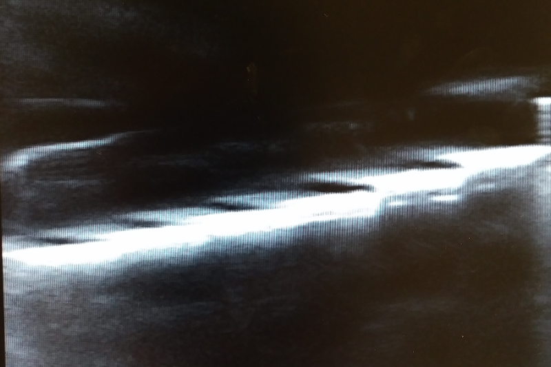
During spinal surgery, surgeons use ball end mills to machine away spinal bone to reach the soft dura underneath. Below this dura are nerves and it is incredibly important that the surgeons not accidentally drill through this. Surgeons currently take MRI scans before the surgery starts to estimate the bone thickness and my research focused on finding alternative methods. I investigated the viability of using ultrasounds or measuring the torque on a drill to see if this was possible. My tests showed that neither of these methods was likely to work which saved us valuable time from pursuing a nonviable project.
